Loading...
Loading...
Loading...
Loading...

Try browsing our complete catalog of products.
Cosmo Bio
Cosmo Bio


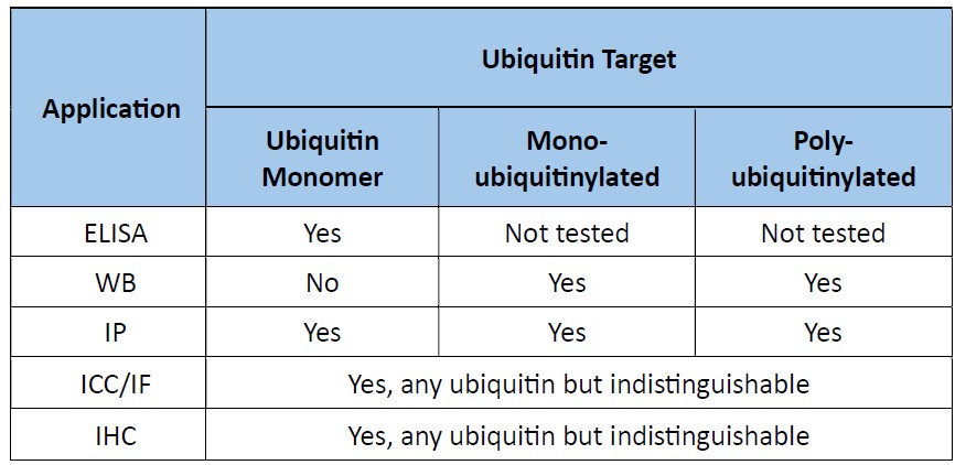
This product is a pan-ubiquitin monobody (single-chain antibody mimic) that recognizes ubiquitin monomers and mono- and poly-ubiquitinated proteins. It boasts several features (described below) that establish its superiority over current pan-ubiquitin monoclonal antibodies such as FK2 and P4D1. It is ideal for immunoprecipitation (IP), immunofluorescence (IF), immunohistochemistry (IHC), immunocytochemistry (ICC), Western blot (WB) and ELISA.
*This product is commercialized under license from the Tokyo Metropolitan Institute of Medical Science, Tokai National University Organization, and Kyoto University.
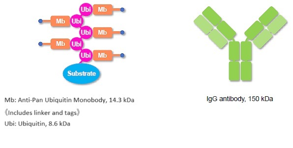
Diagrammatic size comparison – monobody vs IgG
* Monobodies have high specific activity, and better results are obtained at higher dilutions (lower concentrations) than those used with conventional antibody products.
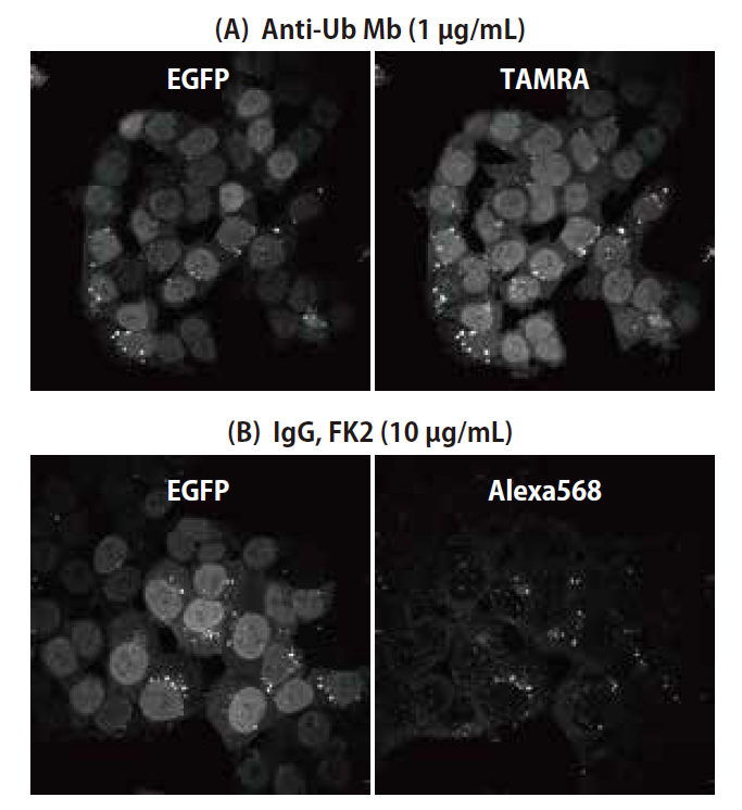
Figure 1. Immunofluorescence microscopy with pan-ubiquitin monobody or pan-ubiquitin mouse monoclonal antibody FK2. HCT-116 cells stably expressing EGFP-ubiquitin fusion protein were treated with puromycin to induce the formation of ubiquitin-positive aggregates, and then treated with (A) TAMRA-labeled pan-ubiquitin monobody or (B) pan-ubiquitin mouse monoclonal antibody FK2 and Alexa 568-labeled secondary antibody. Data was provided by the Tokyo Metropolitan Institute of Medical Science.
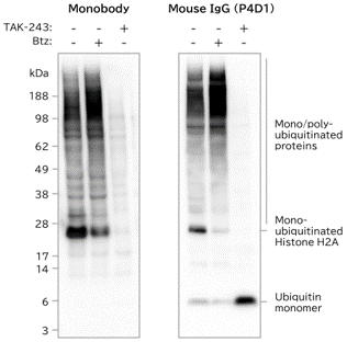
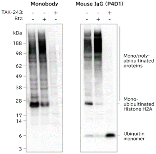
Figure 2. Western blot of ubiquitinated proteins with pan-ubiquitin monobody or pan-ubiquitin mouse monoclonal antibody P4D1. Lysates from HCT-116 cells treated with TAK-243 (inhibitor of Ubiquitin activating enzyme), Btz (proteasome inhibitor) or nothing (-) were Western blotted with pan-ubiquitin monobody (left) or pan-ubiquitin mouse monoclonal antibody P4D1 (right) and visualized using HRP and chemiluminescence. Mono-ubiquitinated histone H2A and small mono/poly-ubiquitinated proteins were detected more strongly with the monobody than with P4D1.
*This product recognizes ubiquitin monomers by ELISA (Figure 6) and immunoprecipitation (Figure 3) but not by Western blotting as shown above in Figure 2.
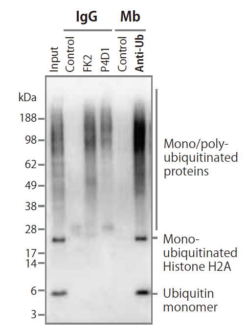
Figure 3. Immunoprecipitation with pan-ubiquitin monobody or pan-ubiquitin mouse monoclonal IgG antibodies FK2 and P4D1. HCT-116 cell extracts were immunoprecipitated with pan-ubiquitin monobody or mouse monoclonal IgG antibodies FK2 and P4D1, followed by Western blotting with P4D1. Only the pan-ubiquitin monobody was able to immunoprecipitate ubiquitin monomers and mono-ubiquitinated Histone H2A.
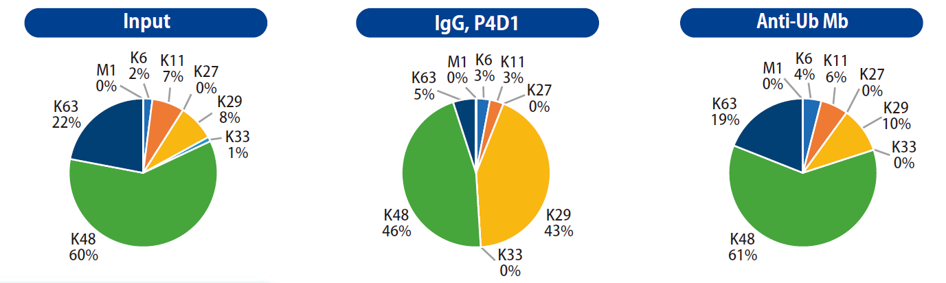
Figure 4. Quantitative mass spectrometry of pan-ubiquitin monobody and monoclonal immunoprecipitates. The immunoprecipitates from Figure 3 (above) 62 kDa or greater were subjected to quantitative mass spectrometry using LC-MS/MS. The quantitative distribution of ubiquitin linkages immunoprecipitated with our monobody closely mirrored that of the input lysate, as compared to the distorted distribution resulting from immunoprecipitation with monoclonal IgG P4D1.
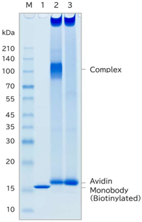
Figure 5. SDS-PAGE analysis of purity and extent of biotinylation. 1 µg of pan-ubiquitin monobody (1), 1 µg of pan-ubiquitin monobody incubated with 5 µg avidin (2), and 5 µg avidin (3) were separated by SDS-PAGE and stained with CBB. The result shows that the monobody is highly pure and biotin-labeled at high yield.
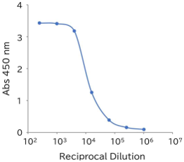
Figure 6. Binding of pan-ubiquitin monobody to immobilized human ubiquitin monomers. Dilution series of pan-ubiquitin monobody (0.5 mg/mL) reacted on an immunoassay plate coated with recombinant human ubiquitin monomer (1 µg/well), followed by secondary reaction with NeutrAvidin-HRP. The result shows the pan-ubiquitin monobody binds with high potency to recombinant human monomeric ubiquitin in ELISA.
| Product Specifications | |
| Application | IP, IHC, ICC, IF, WB |
| Reactivity | All |
| Clonality | Monobody |
| Documents & Links for Anti Pan Ubiquitin Monobody (Biotin labeled) | |
| Datasheet | Anti Pan Ubiquitin Monobody (Biotin labeled) Datasheet v1.1 |
| Flyer | Anti Pan Ubiquitin Monobody (Biotin labeled) Flyer |
| Documents & Links for Anti Pan Ubiquitin Monobody (Biotin labeled) | |
| Datasheet | Anti Pan Ubiquitin Monobody (Biotin labeled) Datasheet v1.1 |
| Flyer | Anti Pan Ubiquitin Monobody (Biotin labeled) Flyer |
This product is a pan-ubiquitin monobody (single-chain antibody mimic) that recognizes ubiquitin monomers and mono- and poly-ubiquitinated proteins. It boasts several features (described below) that establish its superiority over current pan-ubiquitin monoclonal antibodies such as FK2 and P4D1. It is ideal for immunoprecipitation (IP), immunofluorescence (IF), immunohistochemistry (IHC), immunocytochemistry (ICC), Western blot (WB) and ELISA.
*This product is commercialized under license from the Tokyo Metropolitan Institute of Medical Science, Tokai National University Organization, and Kyoto University.
Diagrammatic size comparison – monobody vs IgG
* Monobodies have high specific activity, and better results are obtained at higher dilutions (lower concentrations) than those used with conventional antibody products.
Figure 1. Immunofluorescence microscopy with pan-ubiquitin monobody or pan-ubiquitin mouse monoclonal antibody FK2. HCT-116 cells stably expressing EGFP-ubiquitin fusion protein were treated with puromycin to induce the formation of ubiquitin-positive aggregates, and then treated with (A) TAMRA-labeled pan-ubiquitin monobody or (B) pan-ubiquitin mouse monoclonal antibody FK2 and Alexa 568-labeled secondary antibody. Data was provided by the Tokyo Metropolitan Institute of Medical Science.
Figure 2. Western blot of ubiquitinated proteins with pan-ubiquitin monobody or pan-ubiquitin mouse monoclonal antibody P4D1. Lysates from HCT-116 cells treated with TAK-243 (inhibitor of Ubiquitin activating enzyme), Btz (proteasome inhibitor) or nothing (-) were Western blotted with pan-ubiquitin monobody (left) or pan-ubiquitin mouse monoclonal antibody P4D1 (right) and visualized using HRP and chemiluminescence. Mono-ubiquitinated histone H2A and small mono/poly-ubiquitinated proteins were detected more strongly with the monobody than with P4D1.
*This product recognizes ubiquitin monomers by ELISA (Figure 6) and immunoprecipitation (Figure 3) but not by Western blotting as shown above in Figure 2.
Figure 3. Immunoprecipitation with pan-ubiquitin monobody or pan-ubiquitin mouse monoclonal IgG antibodies FK2 and P4D1. HCT-116 cell extracts were immunoprecipitated with pan-ubiquitin monobody or mouse monoclonal IgG antibodies FK2 and P4D1, followed by Western blotting with P4D1. Only the pan-ubiquitin monobody was able to immunoprecipitate ubiquitin monomers and mono-ubiquitinated Histone H2A.
Figure 4. Quantitative mass spectrometry of pan-ubiquitin monobody and monoclonal immunoprecipitates. The immunoprecipitates from Figure 3 (above) 62 kDa or greater were subjected to quantitative mass spectrometry using LC-MS/MS. The quantitative distribution of ubiquitin linkages immunoprecipitated with our monobody closely mirrored that of the input lysate, as compared to the distorted distribution resulting from immunoprecipitation with monoclonal IgG P4D1.
Figure 5. SDS-PAGE analysis of purity and extent of biotinylation. 1 µg of pan-ubiquitin monobody (1), 1 µg of pan-ubiquitin monobody incubated with 5 µg avidin (2), and 5 µg avidin (3) were separated by SDS-PAGE and stained with CBB. The result shows that the monobody is highly pure and biotin-labeled at high yield.
Figure 6. Binding of pan-ubiquitin monobody to immobilized human ubiquitin monomers. Dilution series of pan-ubiquitin monobody (0.5 mg/mL) reacted on an immunoassay plate coated with recombinant human ubiquitin monomer (1 µg/well), followed by secondary reaction with NeutrAvidin-HRP. The result shows the pan-ubiquitin monobody binds with high potency to recombinant human monomeric ubiquitin in ELISA.
| Product Specifications | |
| Application | IP, IHC, ICC, IF, WB |
| Reactivity | All |
| Clonality | Monobody |
| Documents & Links for Anti Pan Ubiquitin Monobody (Biotin labeled) | |
| Datasheet | Anti Pan Ubiquitin Monobody (Biotin labeled) Datasheet v1.1 |
| Flyer | Anti Pan Ubiquitin Monobody (Biotin labeled) Flyer |
| Documents & Links for Anti Pan Ubiquitin Monobody (Biotin labeled) | |
| Datasheet | Anti Pan Ubiquitin Monobody (Biotin labeled) Datasheet v1.1 |
| Flyer | Anti Pan Ubiquitin Monobody (Biotin labeled) Flyer |
No reviews have been added for this product.